ASP Health performed a fully automated ROSE workflow at Loyola University Medical Center. We combined our automated sample preparation system with a remote telepathology system, so that the cytopathologists could review the slides remotely without having to be present in the Bronchoscopy suite. Dr. Amit Goyal and the cytopathology team at Loyola University led this revolutionary procedure.
The purpose of this procedure was to demonstrate for the first time at Loyola the sample-to-diagnosis benefit of Rapid On-Site Evaluation without the presence of cytology personnel in the operating room. At most sites, ROSE involves the presence of a Cytotechnologist, Cytopathologist, or both. Due to staff shortages or busy schedules, staff is often unavailable, meaning the ROSE procedure cannot be performed. This leaves the Pulmonologist somewhat blind as to whether or not he/she was able to obtain an adequate or diagnostically relevant sample.
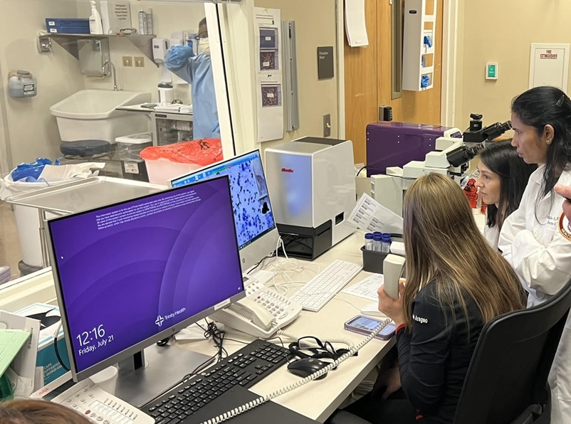
This procedure involved the use of ASP Health’s ROSE Prep™ pro automated sample preparation system and a remote microscopy system demonstrating that ROSE could be performed without valuable cytology resources spent inside the bronchoscopy suite. The ROSE Prep™ pro system was run by a Respiratory Technician in the bronchoscopy suite which automated the deposit and staining process for the samples obtained during the EBUS-FNA procedure. The remote microscopy system that was utilized was provided by Motic and controlled remotely by the Cytopathologist sitting in their office.
The workflow was as follows:
- The sample was obtained using the FNA needle and was directly loaded into ASP’s custom sample vial.
- The sample was mixed with a buffer by massaging the vial to ensure a homogenous sample prior to loading a few (1-4) drops into the ASP ROSE Prep™ pro system.
- The instrument was started with a simple push of a button, while the technician was able to prepare for and collect the next sample from the Pulmonologist.
- Once the sample preparation was complete (< 2 minutes) the slide was placed onto the Motic stage where Dr. Guliz Barkan, Loyola’s Director of Cytopathology, controlled the microscope remotely from her office to review the slide for the presence of diagnostic cells.
- Once identified, she made the final diagnosis and conveyed the information directly to Dr. Amit Goyal, the Interventional Pulmonologist performing the procedure.
This demonstrated for the first time that the ROSE procedure could easily be performed with the ROSE Prep™ pro system and a remote microscopy system without cytopathology personnel in the bronchoscopy suite. This demonstration did have some limitations, as the Motic System was not connected to the high-speed internet of the hospital at the time. This slowed the remote operation of the microscope. However, these issues will be easily resolved in the future with proper network connectivity and better remote desktop sharing software.